lv volume curve | left ventricular pressure volume cv lv volume curve End-diastolic pressure–volume relationship concepts. (A) V30 is the left ventricular (LV) volume at a pressure of 30 mmHg and reflects compliance. A shift to the left suggests diastolic . $12K+
0 · ventricular volume loop diagram
1 · ventricular volume and lvp
2 · ventricular pressure volume loop diagram
3 · ventricular pressure volume curve
4 · left ventricular pressure volume relationship
5 · left ventricular pressure volume ratio
6 · left ventricular pressure volume diagram
7 · left ventricular pressure volume cv
The Lady-Datejust 28 is furnished with either the iconic President’s bracelet – newly optimized with ceramic inserts to prevent stretching out .
ventricular volume loop diagram
chanel reissue 226 vs medium flap
ventricular volume and lvp
To illustrate the pressure-volume relationship for a single cardiac cycle, the cycle can be divided into four basic phases: ventricular filling (phase a, diastole), isovolumetric contraction (phase .EDPVR (End-Diastolic Pressure-Volume Relationship) shows the relationship between ESV and left ventricular volume. The EDPVR curve shows that the left ventricle can withstand large .End-diastolic pressure–volume relationship concepts. (A) V30 is the left ventricular (LV) volume at a pressure of 30 mmHg and reflects compliance. A shift to the left suggests diastolic .
If pulmonary venous flow is decreased, then the ventricle will fill to a smaller end-diastolic volume (decreased preload; green loop in figure). This will cause stroke volume to .A shift of the curve to A indicates that a higher left ventricular (LV) pressure will be required to distend the LV to a similar volume, indicating that the ventricle is less distensible. The slope of .Pressure-volume loops are graphs showing the changing relationship between left ventricular pressure and volume during a cardiac cycle. They provide useful information such as stroke .

As shown in the figure, an increase in afterload shifts the Frank-Starling curve down and to the right (from point A to B), which decreases stroke volume (SV) and increases left ventricular .To illustrate the pressure-volume relationship for a single cardiac cycle, the cycle can be divided into four basic phases: ventricular filling (phase a, diastole), isovolumetric contraction (phase b, systole), ejection (phase c, systole), and isovolumetric relaxation (phase d, diastole).
chanel patent medium boy flap pink
EDPVR (End-Diastolic Pressure-Volume Relationship) shows the relationship between ESV and left ventricular volume. The EDPVR curve shows that the left ventricle can withstand large pressure increases but at a certain threshold, pressure rises rapidly with further volume increases. A PV loop plots the changes in ventricular pressure associated with the changes in volume which occur during the cardiac cycle. Multiple pieces of information can be extracted from such a loop, including volumes (eg. stroke volume), pressures (eg. end-systolic pressure), and area (which represents myocardial work).End-diastolic pressure–volume relationship concepts. (A) V30 is the left ventricular (LV) volume at a pressure of 30 mmHg and reflects compliance. A shift to the left suggests diastolic dysfunction (red), to the right ventricular remodelling (blue). If pulmonary venous flow is decreased, then the ventricle will fill to a smaller end-diastolic volume (decreased preload; green loop in figure). This will cause stroke volume to decrease (from 75 to 50 mL in figure) and EF to decrease from 60 to 50%, but the end-systolic volume will be unchanged.

A shift of the curve to A indicates that a higher left ventricular (LV) pressure will be required to distend the LV to a similar volume, indicating that the ventricle is less distensible. The slope of the LV end-diastolic pressure-volume relation indicates the passive chamber stiffness.Pressure-volume loops are graphs showing the changing relationship between left ventricular pressure and volume during a cardiac cycle. They provide useful information such as stroke volume or end-diastolic volume, as well as systolic, diastolic, and pulse pressure.As shown in the figure, an increase in afterload shifts the Frank-Starling curve down and to the right (from point A to B), which decreases stroke volume (SV) and increases left ventricular end-diastolic pressure (LVEDP). The basis for this is found in .
Background: Left ventricular (LV) efficiency is a key pathophysiological marker in heart failure (HF). LV pressure curve estimation and three-dimensional (3D) volumes via echocardiography allows for non-invasive pressure-volume (PV) analysis and calculation of an index of efficiency (Figure 1). Aim: Validate efficiency index by 3D echocardiography by .
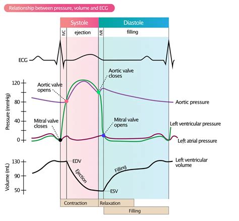
This pressure volume loop diagram represents the 5 phases of the cardiac cycle. It makes more sense to start at the end of stage 4 of the cardiac cycle: isovolumetric relaxation and beginning of stage 5- ventricular diastole. At this point, the end systolic volume of the LV is +- .
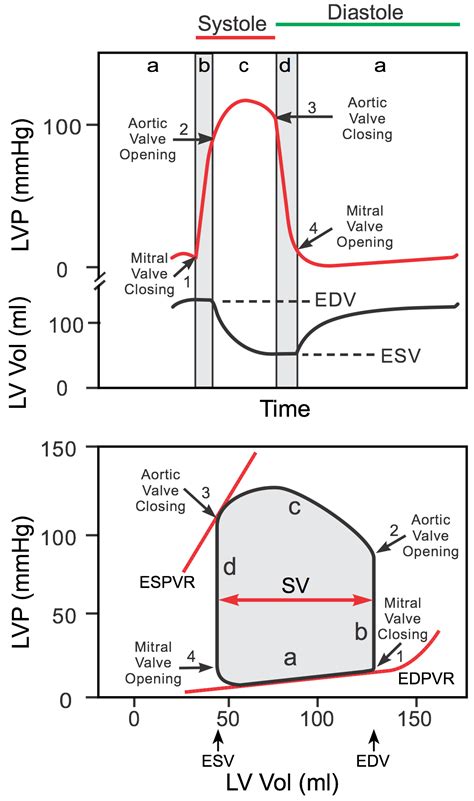
To illustrate the pressure-volume relationship for a single cardiac cycle, the cycle can be divided into four basic phases: ventricular filling (phase a, diastole), isovolumetric contraction (phase b, systole), ejection (phase c, systole), and isovolumetric relaxation (phase d, diastole).
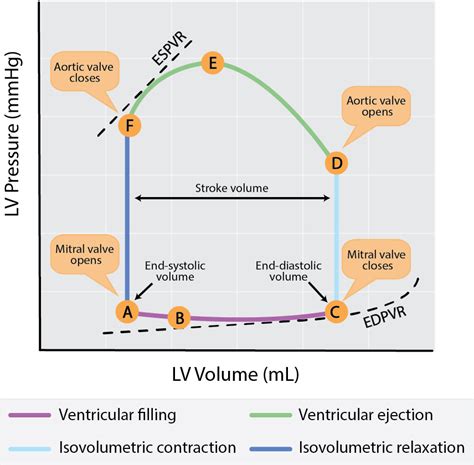
EDPVR (End-Diastolic Pressure-Volume Relationship) shows the relationship between ESV and left ventricular volume. The EDPVR curve shows that the left ventricle can withstand large pressure increases but at a certain threshold, pressure rises rapidly with further volume increases.
A PV loop plots the changes in ventricular pressure associated with the changes in volume which occur during the cardiac cycle. Multiple pieces of information can be extracted from such a loop, including volumes (eg. stroke volume), pressures (eg. end-systolic pressure), and area (which represents myocardial work).End-diastolic pressure–volume relationship concepts. (A) V30 is the left ventricular (LV) volume at a pressure of 30 mmHg and reflects compliance. A shift to the left suggests diastolic dysfunction (red), to the right ventricular remodelling (blue). If pulmonary venous flow is decreased, then the ventricle will fill to a smaller end-diastolic volume (decreased preload; green loop in figure). This will cause stroke volume to decrease (from 75 to 50 mL in figure) and EF to decrease from 60 to 50%, but the end-systolic volume will be unchanged.A shift of the curve to A indicates that a higher left ventricular (LV) pressure will be required to distend the LV to a similar volume, indicating that the ventricle is less distensible. The slope of the LV end-diastolic pressure-volume relation indicates the passive chamber stiffness.
Pressure-volume loops are graphs showing the changing relationship between left ventricular pressure and volume during a cardiac cycle. They provide useful information such as stroke volume or end-diastolic volume, as well as systolic, diastolic, and pulse pressure.
As shown in the figure, an increase in afterload shifts the Frank-Starling curve down and to the right (from point A to B), which decreases stroke volume (SV) and increases left ventricular end-diastolic pressure (LVEDP). The basis for this is found in . Background: Left ventricular (LV) efficiency is a key pathophysiological marker in heart failure (HF). LV pressure curve estimation and three-dimensional (3D) volumes via echocardiography allows for non-invasive pressure-volume (PV) analysis and calculation of an index of efficiency (Figure 1). Aim: Validate efficiency index by 3D echocardiography by .

Published on 11/18/20. Malta, or the Republica of Malta, is an island nation in the central Mediterranean, located between Sicily and North Africa. It includes the main island of Malta (home to the capital city, Valletta), the smaller island of Gozo, and the tiny, barely populated island of Comino.
lv volume curve|left ventricular pressure volume cv


