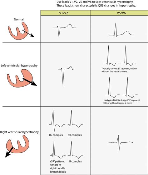
lv ecg | left ventricular hypertrophy life in the fast lane lv ecg ECG Pearl. There are no universally accepted criteria for diagnosing RVH in . Location: Featured Servers. Show your server here. Increase your server visibility using the highlighting service. Boost a server. Servers list. No server matches the search criteria used. Monitoring, stats, banner and more for your game server. Generate your own live signature of your server.
0 · signs of lvh on ecg
1 · lvh ecg pattern
2 · lvh ecg chart
3 · left ventricular hypertrophy life in the fast lane
4 · ecg showing lvh
5 · ecg lvh meaning
6 · ecg findings in lvh
7 · diagnosis of lvh on ecg
RTLinux. RTLinux is a hard realtime real-time operating system (RTOS) microkernel that runs the entire Linux operating system as a fully preemptive process. The hard real-time property makes it possible to control robots, data acquisition systems, manufacturing plants, and other time-sensitive instruments and machines from RTLinux applications.
The left ventricle hypertrophies in response to pressure overload secondary to conditions such as aortic stenosis and hypertension. This results in increased R wave amplitude in the left-sided ECG leads (I, aVL and V4-6) and increased S wave depth in the right-sided .R Wave Peak Time Rwpt - Left Ventricular Hypertrophy (LVH) • LITFL • ECG .ECG Pearl. There are no universally accepted criteria for diagnosing RVH in .ECG Criteria for Left Atrial Enlargement. LAE produces a broad, bifid P wave in .
In LBBB, conduction delay means that impulses travel first via the right bundle .
U Waves - Left Ventricular Hypertrophy (LVH) • LITFL • ECG Library DiagnosisLeft Axis Deviation - Left Ventricular Hypertrophy (LVH) • LITFL • ECG .
The most common causes of left ventricular hypertrophy are aortic stenosis, aortic regurgitation, hypertension, cardiomyopathy and coarctation of the aorta. There are several ECG indexes, . ECG features suggestive of LV dilation (dilated cardiomyopathy) LVH is a bit of a misnomer because this actually reflects increased left ventricular mass (due to increased wall .
signs of lvh on ecg
The left ventricle hypertrophies in response to pressure overload secondary to conditions such as aortic stenosis and hypertension. This results in increased R wave amplitude in the left-sided ECG leads (I, aVL and V4-6) and increased S .The most common causes of left ventricular hypertrophy are aortic stenosis, aortic regurgitation, hypertension, cardiomyopathy and coarctation of the aorta. There are several ECG indexes, which generally have high diagnostic specificity but low sensitivity. ECG features suggestive of LV dilation (dilated cardiomyopathy) LVH is a bit of a misnomer because this actually reflects increased left ventricular mass (due to increased wall thickness or chamber dilation). The following features may suggest LV chamber dilation: Reduction in R-wave of left precordial leads.
Left ventricular hypertrophy, especially in patients with hypertension, increases the risk of heart failure, ischemic heart disease, sudden death, atrial fibrillation and stroke.
Left ventricular hypertrophy (LVH) refers to an increase in the size of myocardial fibers in the main cardiac pumping chamber. Such hypertrophy is usually the response to a chronic pressure or volume load. The two most common pressure overload states are systemic hypertension and aortic stenosis.

According to the American Society of Echocardiography and/European Association of Cardiovascular Imaging, LVH is defined as an increased left ventricular mass index (LVMI) to greater than 95 g/m in women and increased LVMI to greater than 115 g/m in men.Left ventricular hypertrophy can be diagnosed on ECG with good specificity. When the myocardium is hypertrophied, there is a larger mass of myocardium for electrical activation to pass.The traditional approach to the ECG diagnosis of left ventricular hypertrophy (LVH) is focused on the best estimation of left ventricular mass (LVM) i.e. finding ECG criteria that agree with LVM as detected by imaging. The principal ECG‐derived diagnostic characteristics for LVH detection include an increased QRS complex amplitude, an increased QRS complex duration, a leftward shift of electrical axis of the QRS in frontal plane, and ST‐segment deviation and T wave changes associated with left ventricular strain. 9 Furthermore, more advanced ECG methods .
The ECG changes in a patient with left ventricular hypertrophy (LVH) were described 117 years ago by Einthoven in 1906 (Einthoven, 1957). He drew attention to the distinctive finding—the increased QRS amplitude in the “left hand to left foot lead” (i.e., lead III). The left ventricle hypertrophies in response to pressure overload secondary to conditions such as aortic stenosis and hypertension. This results in increased R wave amplitude in the left-sided ECG leads (I, aVL and V4-6) and increased S .The most common causes of left ventricular hypertrophy are aortic stenosis, aortic regurgitation, hypertension, cardiomyopathy and coarctation of the aorta. There are several ECG indexes, which generally have high diagnostic specificity but low sensitivity. ECG features suggestive of LV dilation (dilated cardiomyopathy) LVH is a bit of a misnomer because this actually reflects increased left ventricular mass (due to increased wall thickness or chamber dilation). The following features may suggest LV chamber dilation: Reduction in R-wave of left precordial leads.
Left ventricular hypertrophy, especially in patients with hypertension, increases the risk of heart failure, ischemic heart disease, sudden death, atrial fibrillation and stroke. Left ventricular hypertrophy (LVH) refers to an increase in the size of myocardial fibers in the main cardiac pumping chamber. Such hypertrophy is usually the response to a chronic pressure or volume load. The two most common pressure overload states are systemic hypertension and aortic stenosis. According to the American Society of Echocardiography and/European Association of Cardiovascular Imaging, LVH is defined as an increased left ventricular mass index (LVMI) to greater than 95 g/m in women and increased LVMI to greater than 115 g/m in men.Left ventricular hypertrophy can be diagnosed on ECG with good specificity. When the myocardium is hypertrophied, there is a larger mass of myocardium for electrical activation to pass.
The traditional approach to the ECG diagnosis of left ventricular hypertrophy (LVH) is focused on the best estimation of left ventricular mass (LVM) i.e. finding ECG criteria that agree with LVM as detected by imaging.
The principal ECG‐derived diagnostic characteristics for LVH detection include an increased QRS complex amplitude, an increased QRS complex duration, a leftward shift of electrical axis of the QRS in frontal plane, and ST‐segment deviation and T wave changes associated with left ventricular strain. 9 Furthermore, more advanced ECG methods .
ysl lipstick number 29

ysl lipstick plum fusion
You can verify a Louis Vuitton bag and see if it’s real or fake by checking the “LOUIS VUITTON ®” logo. Fake bags always have thicker text than authentic ones. Related: Legit check ANY Louis Vuitton bag. 1. Interior labels. Every LV bag has this texton the interior label: ® LOUIS VUITTON made in *country’s name*. 1.1. Square label.
lv ecg|left ventricular hypertrophy life in the fast lane


